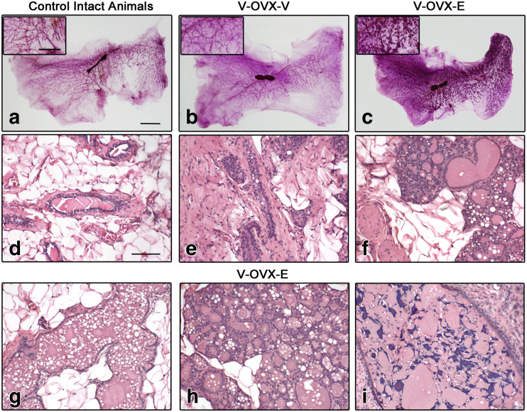
Fig. 2.
ERT in middle-aged OVX rats. Representative images of whole mounts (a–c) and H&E-stained sections (d–i) of intact control animals (a, d), V-OVX-V (b, e), and V-OVX-E rats (c, f–i). The higher lobular development in estrogen-treated rats (c) compared with those given vehicle (b) is noteworthy. In V-OVX-V animals, atrophy of the mammary gland after OVX is characterized by scarce number of ducts and lobuloalveolar structures and, a relative increase in collagen amount (e) compared with intact animals (d). E treatment induced alveolar and ductal dilation (f, g), areas with secretion, vacuolation, and intraepithelial fat globules mimicking secretory activity, as well as lobular hyperplasia (h) and cysts containing small corpora amylacea (i). In the insets, an area between the nipple and the lymph node in each whole mount is magnified. The bar represents the magnification in each set of images: 10 mm for a–c; 5 mm for whole mount insets, and 100 μm for d–i

