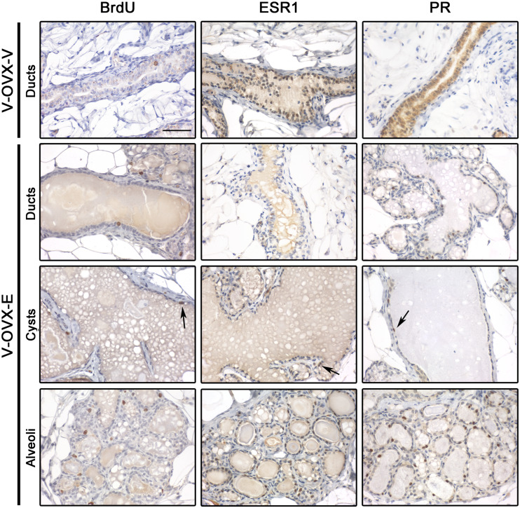
Fig. 3.
Proliferation and steroid hormone receptor expression in V-OVX-V and V-OVX-E rats. All three markers were analyzed in the epithelial cells of ducts, cysts, and alveoli when the structure was present in mammary gland samples. BrdU and ESR1 expression was identified in the nuclei of epithelial cells, regardless of the mammary structure and experimental group. PR expression in V-OVX-V animals was mainly observed in the cytoplasm whereas that in V-OVX-E rats was mainly observed in the nucleus. Positive cells are indicated by the arrows; note the lower ESR1 and PR expression in cysts compared with ducts and alveoli. All images have the same magnification, bar: 100 μm

