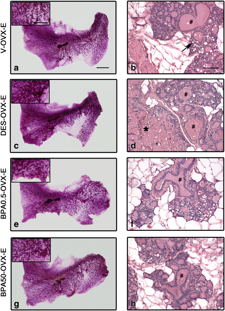
Fig. 4.
ERT in rats perinatally exposed to DES and BPA. Representative images of whole mounts (a, c, e, g) and H&E-stained sections (b, d, f, h) in V-OVX-E (a, b), DES-OVX-E (c, d), BPA0.5-OVX-E (e, f), and BPA50-OVX-E (g, h) treated rats. Perinatal exposure to DES prior to ERT induced greater development of the mammary gland and an increased percentage of ductal cysts compared with non-perinatally exposed rats. In BPA-exposed animals, presence of secretion, dilation of ducts and alveoli, and lobular hyperplasia are observed. In the insets, an area between the nipple and the lymph node in each whole mount is magnified. Dilated ducts (#), alveolar cells with lipid droplets (arrow), and cysts (*) are indicated in the images. The bar represents the magnification in each set of images: 10 mm for a, c, e, and g; 5 mm for whole mount insets; and 100 μm for b, d, f, and h

