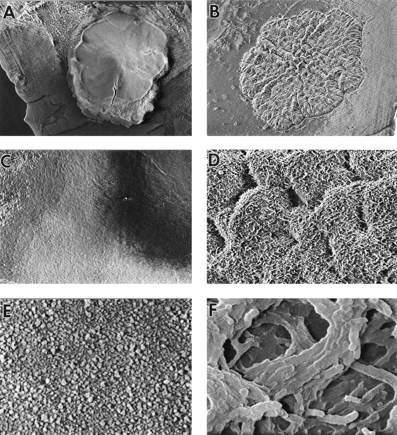
FIG. 7.
Cryo scanning electron micrographs of a wild-type colony (A, C, and E) and a flat colony variant (B, D, and F). (A and B) Colonies viewed from above at a magnification of ×15; (C and D) surfaces of colonies at a magnification of ×120; (E and F) surfaces of colonies at a magnification of ×3,750.

