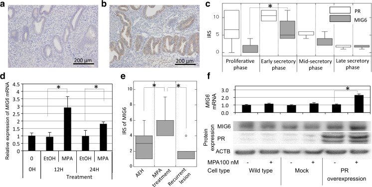
Fig. 1.
a, b Immunohistochemical expression of MIG6 in the normal endometrium in the proliferative phase (a) and secretory phase (b). Positive staining was observed in the secretory phase. c Graphical demonstration of the expression of the progesterone receptor (PR) and MIG6 in the proliferative phase, early, mid, and late secretory phases. Data are presented in the box plot as the median and 25–75 percentile of the IRS. MIG6 expression was stronger in the early to mid-secretory phases when PR was positive. d Relative expression of MIG6 mRNA in cultured normal endometrial glandular (NEG) cells after the addition of MPA at 100 nM. Data are presented as the mean ± SD of three independent experiments. e The immunohistochemical expression of MIG6 in atypical endometrial hyperplasia (AEH), in AEH after the MPA treatment, and in recurrent tumors after the MPA treatment. The box plot indicates median and 25–75 percentile. f The expression of MIG6 mRNA, MIG6, and PR protein in Ishikawa cells transfected with PR (PR overexpression) or empty vector (mock) with or without MPA. The cells were harvested at 24 h after addition of MPA or vehicle. *P < 0.01, IRS immunoreactive score, which was calculated by multiplying the quantity score (no staining as 0, 1–10% as 1, 11–50% as 2, 51–80% as 3, and 81–100% as 4) and staining intensity score (negative as 0, weak as 1, moderate as 2, and strong as 3)

