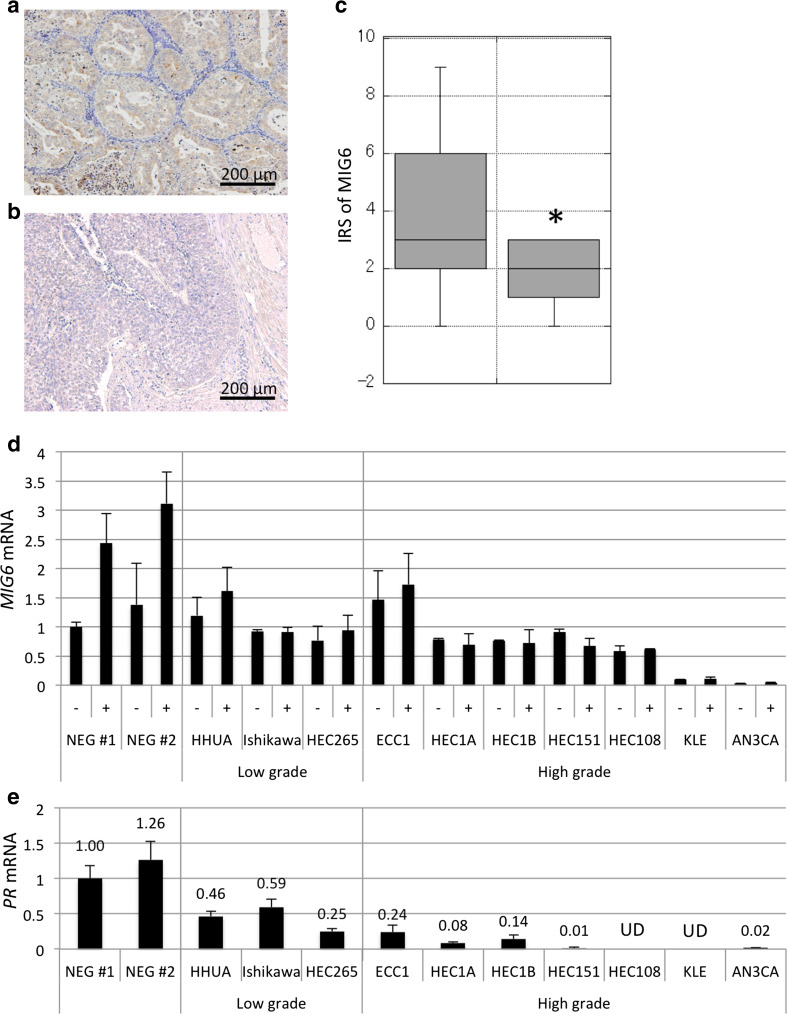
Fig. 2.
a–c Results of immunostaining for MIG6 in grade 1 (a) and grade 3 (b) endometrial carcinomas and its graphical demonstration (the box plot indicating median and 25–75 percentile) (c). The expression of MIG6 decreased in poorly differentiated tumors. d, e The results of real-time PCR indicating the expression of MIG6 (d) and PR (e) mRNA in two NEG cells (NEG no.1 and 2) and various endometrial carcinoma cell lines before and at 24 h after the addition of MPA. The expression of MIG6 was increased by MPA in NEG and HHUA cells but was absent in other cell lines. *P < 0.01, IRS immunoreactive score

