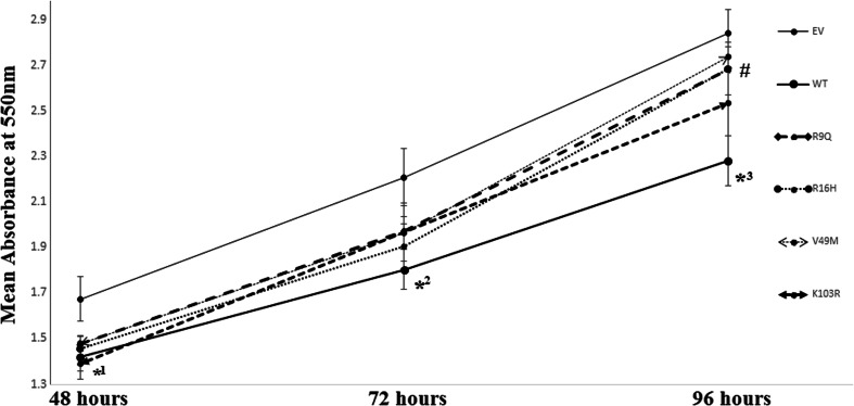
Fig. 1.
Proliferation assays of WT and N-terminus variants of AIP in GH3 cells. GH3 cells were transfected with either pcDNA3 empty vector (EV) or WT or N-terminus variants, and MTT analysis was carried out at 3 time points after transfection. Points represent means of four separate experiments with five replicates per experiment. Error bars indicate standard error. (*1, p = <0.05 comparing all AIP expressing plasmids to EV at 48 h; *2, p = <0.05 comparing all AIP expressing plasmids to EV except the V49M variant at 72 h; *3, p = <0.05 comparing WT-AIP to EV at 96 h; #, p = <0.05 comparing the R9Q, R16H and V49M N-terminus variant to WT-AIP at 48 h)

