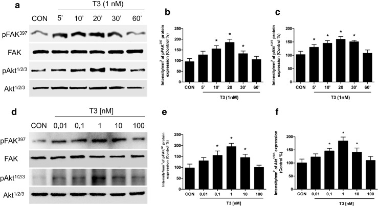
Fig. 2.

T3 induces FAK and Akt phosphorylation/activation in T-47D breast cancer cells. Time (a) and dose-dependent (d) FAK and Akt activation of T-47D cells after T3 treatment. Total cell amount of wild-type FAK and Akt or Tyr397-phosphorylated FAK (p-FAK) and phosphorylated-Akt (p-Akt) are shown by Western blot analysis. Phospho-FAK397 and p-Akt densitometry values were adjusted to FAK and Akt intensity, respectively, and then normalized to the control sample. * = P < 0.05 vs. corresponding control. All experiments were performed in triplicate with consistent results; representative images are shown (b, c, e, f)
