
Figure 3. Prolylcarboxylpeptidase localizes in lysosomes and mediates intracardiac angiotensin II degradation through the lysosomal pathway.
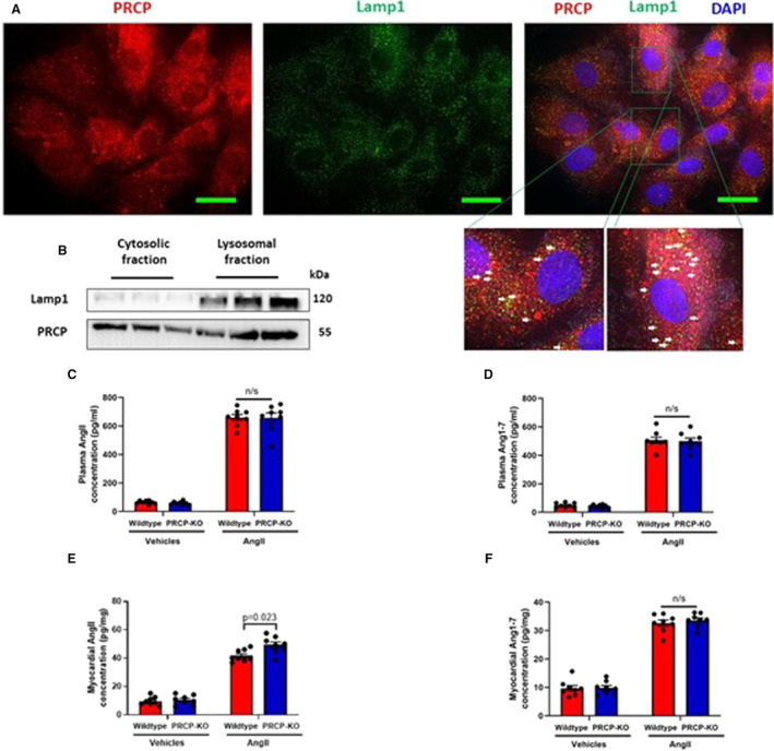
A, Immunostaining of prolylcarboxylpeptidase (red) and Lamp1 (lysosome marker) in H9C2 cells shows prolylcarboxylpeptidase partially resides in lysosomes. White arrows indicate colocalization of prolylcarboxylpeptidase and Lamp1 (yellow dots) (scale bar: 15 μm). B, Cytosolic and lysosomal fractions of wild‐type hearts show the localization of prolylcarboxylpeptidase in both cytosol and lysosome. Lamp1 is used as a marker for lysosomes. C, Angiotensin II levels in plasma. D, Angiotensin 1‐7 levels in plasma. E, Angiotensin II levels in myocardial tissues. F, Angiotensin 1‐7 levels in myocardial tissues (n=8 mice per group). Data are expressed as means±SEM. Two‐way ANOVA with Bonferroni correction for post hoc comparisons was used for analysis. AngII indicates angiotensin II; DAPI, 4′, 6‐diamidino‐2‐phenylindole; Lamp1, lysosomal‐associated membrane protein 1; PRCP, prolylcarboxylpeptidase; and PRCP‐KO, prolylcarboxylpeptidase knockout.