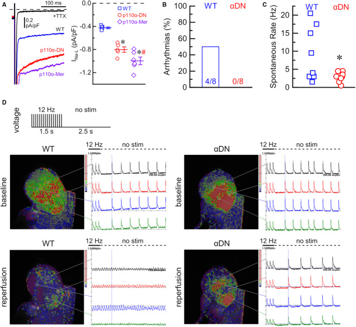
Figure 5. Late Na+ current and reperfusion arrhythmias in PI3Kα mutant hearts.

A, Left: representative late Na+ currents (INa‐L) in response to step depolarization from −120 to −40 mV in wild type (WT, blue), constituent PI3Kα‐deficient (p110α‐DN, red), and inducible PI3Kα‐deficient (p110α‐Mer, purple) myocytes, and background current after application of specific Na+ channel blocker, tetrodotoxin (TTX) in black; Right: average current densities of INa‐L (TTX‐sensitive current) for WT (blue), p110α‐DN (red), and p110α‐Mer (purple) myocytes. B, Fraction of arrhythmias at reperfusion in wild‐type (WT) and p110α‐DN (αDN) excised hearts. C, Spontaneous heart rate (no stimulation) after reperfusion in WT and p110α‐DN (αDN) excised hearts. D, Representative images and voltage traces from WT and p110α‐DN (αDN) excised hearts at baseline and reperfusion; no stim, no stimulation. Data are presented as mean±SEM; statistical significance is calculated using 1‐way ANOVA in (A) and 2‐sided t test with Welsch correction in (C); *P<0.05 vs WT (A, C), # P<0.05 vs p110α‐DN (A).
