Figure 2.
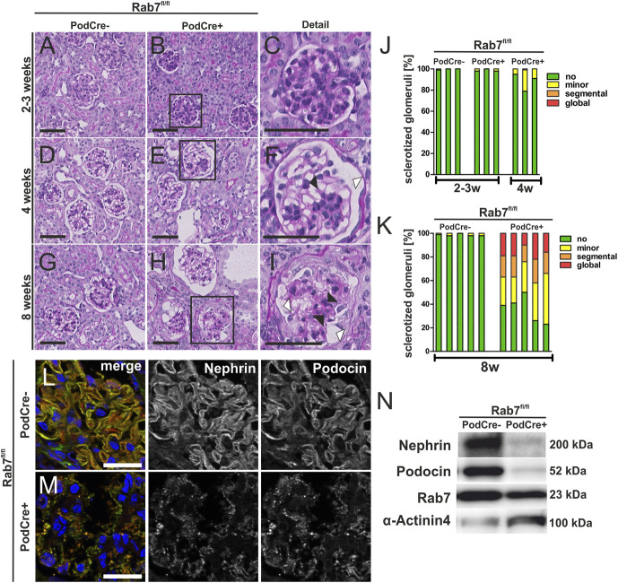
Rab7 depletion of podocytes leads to FSGS. (A–I) PAS staining of Rab7fl/fl;PodCre+ mice at age 2–3, 4, and 8 weeks. Black arrowheads mark sclerosis of the glomerulus and Bowman capsule; white arrowheads mark the dilated Bowman space, scale bar: 50 nm. (J and K) Glomerulosclerosis score of 2–3-week-old and 4-week-old (J) and 8-week-old mice (K) (data are shown in %). Thresholds of sclerotization were defined as following: no alteration; minor, mesangial expansion and/or thickening of the basement membrane without sclerosis; segmental, ≤50%; and global, >50% tuft area of each glomerulus affected. Three (J) or five (K) animals per group and >50 glomeruli per animal were analyzed. An unpaired test with Welch correction was performed for 8-week-old control versus knock out (KO) mice with a significance of P = 0.0002. (L/M) Immunofluorescence staining of the SD proteins nephrin and podocin at age 4 weeks, scale bar: 50 µm. (N) Western blot analysis of glomerular lysates for the slit diaphragm proteins nephrin and podocin in 4-week-old mice show decreased nephrin and podocin levels. Rab7 is still detectable since Rab7 is only depleted in podocytes, and whole glomerular lysates were used. KO, knock out; SD, slit diaphragm. Figure 2 can be viewed in color online at www.jasn.org.

