Figure 5.

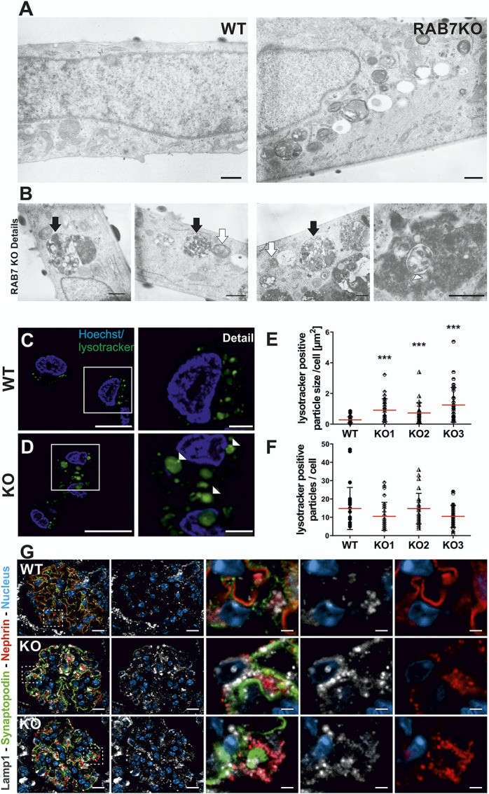
Rab7 depletion in podocytes results in accumulation of enlarged LAMP2-positive vesicles. (A) Ultrastructural analysis of Rab7-depleted podocytes shows different electron-dense structures accumulating compared with the wildtype (overview). Scale bar: 500 nm. (B) Detailed ultrastructural analysis of Rab7-depleted podocytes focusing on three accumulating vesicle types: black arrow, large single-membrane vesicle containing multiple intraluminal vesicles; white arrow, smaller, single-membrane vesicles containing membrane layers, and occasionally found double-membrane vesicles (white arrowhead). Scale bar: 500 nm. (C/D) Live cell staining with LysoTracker green revealed enlarged acidic compartments in Rab7-depleted cells (white arrowheads). (E/F) Quantification of the size and number of LysoTracker-positive particles in Rab7-depleted cell lines. ***P < 0.0001 by the unpaired two-tailed Mann–Whitney test, shown are mean with SD; cells analyzed: wildtype (wt): N=112, Rab7 knock out [knock out]: N=84. (G) Immunofluorescence analyses with control (wild type [WT], Rab7fl/fl; PodCre−) and Rab7fl/fl; PodCre+ (KO) mice. Images show representative IF-staining against Lamp1 (white) in glomeruli of WT and KO animals at 8 weeks. Nephrin (red), synaptopodin (green), and nuclei (Dapi, blue) were stained as comarkers. Nephrin-labeling shows a vesicular-like pattern and translocation toward the podocyte cell body apical of synaptopodin in KO animals. Synaptopodin shows intact localization to the basolateral podocyte compartment. Analysis of Lamp1 revealed enrichment of Lamp1-positive vesicles in the podocyte compartment of KO animals and colabeling of most but not all nephrin-positive structures. Interestingly, a subset of Lamp1-positive vesicles with perinuclear localization is negative for nephrin. Scale bar: overview=10 µm, details=2 µm. SD, slit diaphragm; WT, wild type. Figure 5 can be viewed in color online at www.jasn.org.
