Figure 5.
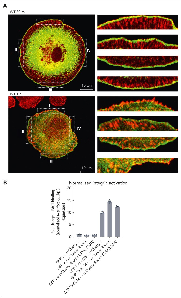
Cellular location of filamin and its regulation on integrin-ECM ligand binding and wound healing. (A) Filamin localizes to nascent FAs upon integrin activation. Merged confocal sections of EGFP (enhanced green fluorescent protein)-filamin WT were fixed after plating on FN-coated glass for 30 minutes or 1 hour. Four randomly selected regions (dotted boxes labeled I-IV, left panel) were enlarged (right) to show the colocalization at the cell cortex. The enlarged boxes on the right correspond as from I to IV from top to bottom, respectively. Note that the resolving power of a confocal microscope is, on average, from 100 to 200 nm laterally (x-y–axes), and 500 nm axially (z-axis). Given that α/β CTs are separated by >100Å61 and that extended forms of both filamin26 and talin63 are >100 nm, filamin (bound to α CT) and vinculin (bound to talin-β CT) can be easily separated beyond the resolving power of a confocal microscope. Scale, 10 μm; green, EGFP-filamin; red, vinculin. Green fluorescent protein–expressing cells were considered filamin-transfected and therefore selected for imaging. See supplemental Figure 4 for individual stains. (B) Filamin promotes active talin-triggered integrin activation. Bar graph generated from flow cytometry data using PAC-1 antibody to analyze the level of activated integrins on cells transfected with indicated vectors. Data are averages ± SEM for 3 independent experiments.

