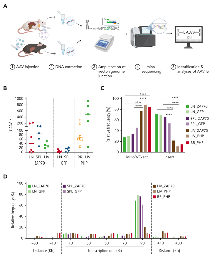
Figure 1.
Retrieval of AAV ISs. (A) Schema of the experimental strategy. LN cells, SPL, and LIV tissues were collected from Zap70-deficient mice treated with AAV8. BR and LIV tissues were collected from Mecp2-deficient mice systemically injected with AAV-PHP. DNA was purified from the collected tissues and SLiM-PCR was performed to amplify vector/host-genome junction. PCR amplicons were assembled into libraries, sequenced, and reads were analyzed using RAAVIoli, a bioinformatics pipeline tailored to identify AAV ISs. (B) The numbers of AAV ISs retrieved from LN-derived mature T cells from LN, SPL, LIV, and BR tissues from the different groups of treated mice are presented with the median indicated as a colored line. (C) Relative frequency (indicated as percentage) of AAV ISs characterized by either precise homology breakpoints (Exact) and microhomology regions (MHoRs) between the vector and the host chromosomal sequences or random nucleotide insertions (insert). (D) Integration site distribution within gene bodies and their surrounding genomic regions. Each gene interval was quantified from the TSS up to the end of its coding region; this interval is considered as 100% and then normalized in bins of 20%. The surrounding genomic regions are divided in intervals of 20 kb. In panel C, statistical analyses were performed using a Fisher exact test ∗∗∗∗ P < .0001; see supplemental Table 2 for detailed statistical comparison.

