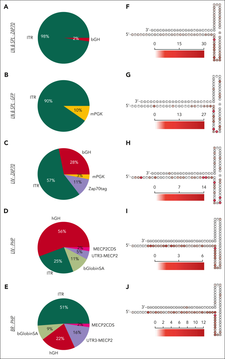
Figure 5.
AAV genomic features at the vector/host-genome junction. (A-E) Pie charts indicating the frequency of AAV features found at the junction breakpoint between the integrated vector and the host genomic region for the different IS data sets as indicated. (F-J) Heatmap of the AAV 3′ITR secondary structure with the red scale indicating the frequency of AAV insertions occurring at the indicated nucleotide position for the different IS data sets. bGlobinSA, beta globin splice acceptor; CDS, coding sequence; hGH, human growth hormone; mPGK, murine phosphoglycerate kinase.

