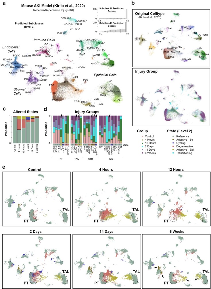
Extended Data Fig. 6. Altered states in a mouse model of AKI.
a. UMAP showing mouse AKI (IRI) data4 with cell types predicted from snCv3. Mouse datasets were projected onto the snCv3 UMAP embeddings (Fig. 2b). Histograms of prediction scores for subclasses (level 1 and 3) are shown. b. UMAP plots as in (a) showing the original cell type annotations4 and injury groups (time points following IRI) for mouse data. c. Barplot showing the proportion of altered states for each mouse injury group. d. Barplot showing proportion of each injury group for a subset of predicted subclasses. Arrows indicate altered states or immune cells (MAC-M2) that persisted at 6 weeks following injury. e. UMAP as in (a) showing the distribution of reference and altered states over the different injury groups.

