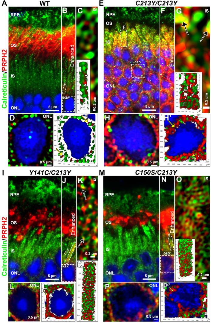
Fig. 7.
C213Y/C213Y retinas show PRPH2 in the ER and inner segment. P30 retinal cross sections of WT and C213Y-containing mutant retinal sections were labeled for PRPH2 (red) and the ER marker calreticulin (green). B, F, J, and N are portions of images shown in A, E, I, and M (respectively) in which the PRPH2 signal intensity was increased past saturation of the outer segment in order to visualize the small amount of PRPH2 in the inner segment. F is a enhance portion of the image in E, and was included for comparison only. C, G, K, K′ and O are images (or 3D renders) of PRPH2 and calreticulin labeling in the inner segment region marked in the adjacent panel. Images were captured using high-resolution Airyscan confocal imaging. Black arrows point to areas of colocalization. D, D′, H, H′, L, L′, P, and P′ are images of perinuclear localization of PRPH2 and calreticulin captured using Airyscan. The images were expanded from the regions marked in the panel above. 3D renders were generated from high-resolution images. Yellow arrows point to perinuclear localization of PRPH2. Scale bar is 5 µm in A, E, I, and M, 0.2 µm in C, G, K, and O, and N 0.5 µm in D, H, L, and P. RPE retinal pigment epithelium, OS outer segment, IS inner segment, ONL outer nuclear layer

