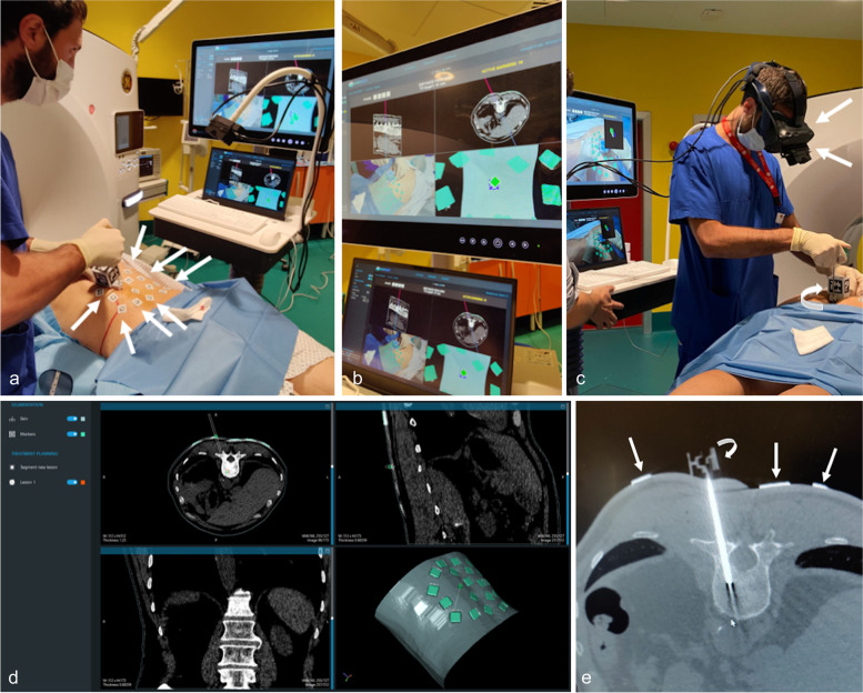
Fig. 1.
A representative case with lumbar bone lesion from our study population. The fiducial markers are placed on patient’s skin around the entry point of percutaneous biopsy (arrows in a and e). The AR system (Endosight, R.A.W. Srl, Milan, Italy) consists of a 27″ medical display (ACL, Leipzig, Germany), a laptop (Dell Technologies, Round Rock, TX, USA) with AR software (b), and a commercially available head-mounted display (HMD) (Oculus Rift-S, Facebook Technologies, Menlo Park, CA, USA) paired with a binocular camera (Zed Mini, Stereolabs, San Francisco, CA, USA) (arrows in c) that is used to coregister the fiducial markers segmented on CT images with those positioned to the patient’s skin (b). The best needle trajectory to reach the target is built (d). A specific marker attached to the needle to monitor its position and angle during the procedure (curved arrows in c and e)

