Fig. 2.
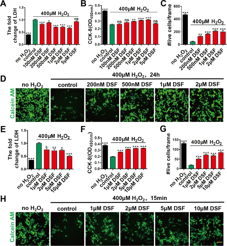
DSF treatment protects CMs from H2O2-induced injury in a dose-dependent manner. A-D Fold change of LDH release (A), CCK-8 detection (B), numbers of live CMs (C), and representative images of calcein AM staining (D) in NRVMs after exposure to H2O2 (400 μM) and different concentrations of DSF for 24 h. n = 6 (A, B), or n = 5 (C, D) per group. E–H Fold change of LDH release (E), CCK-8 detection (F), numbers of live CMs (G), and representative images of calcein AM staining (H) in NRVMs exposed to 400 μM H2O2 for 15 min, followed by treatment with different concentrations of DSF for 24 h. n = 6 (E, F), or 5 (G, H) per group. Scale bar, 100 μm (D, H). *p < 0.1, **p < 0.01, ***p < 0.001; ns, no significant difference. One-way ANOVA with Dunnett’s test (A-C, E–G). Data are the mean ± s.e.m. (A-C, E–G)

