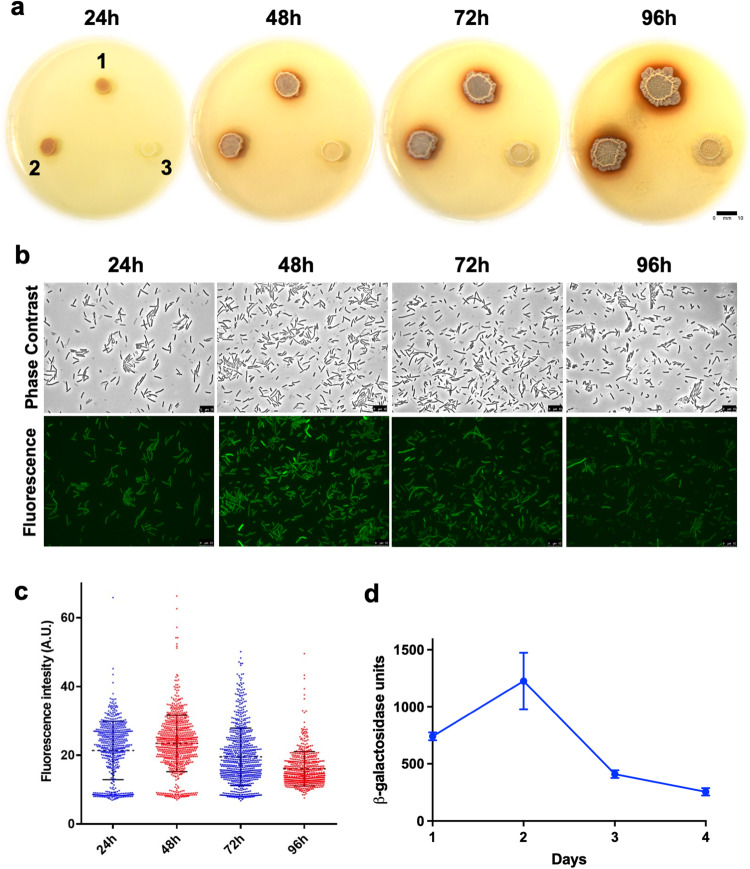
Fig. 1. Strong production of pulcherrimin and activities of its biosynthetic genes during B. subtilis biofilm development.
a Colony biofilms of the B. subtilis wild-type strain (3610, 1), the complementation strain (LA33, 2), and the pulcherrimin biosynthetic mutant ΔyvmC-cypX (YC800, 3) grown on biofilm-inducing media LBGM supplemented with 0.2 mM FeCl3. Plates were incubated at 30 °C and images were taken every 24 h over the course of 4 days. Scale bar, 10 mm. b Induction of the pulcherrimin biosynthesis operon during biofilm formation using the reporter strain bearing the transcriptional fusion PyvmC-gfp (LA20). Pellicle biofilms developed in LBGM and incubated statically at 30 °C. Pellicles were then harvested and cells examined under fluorescence microscopy every 24 hours over the course of 4 days. Representative phase and fluorescence images from each time point were shown. Scale bar, 10 μm. c Violin plots demonstrating the distribution of fluorescence expressed from wild-type cells bearing the PyvmC-gfp reporter (LA20). Cells were collected at 4 different time points during pellicle biofilm development (from 24 h to 96 h, as shown in b above). Each dot represents a single cell. Fluorescence pixel quantification of cells was carried out by using the MicrobeJ. Median values are represented by dashed horizontal lines. Solid lines represent standard deviation. d Activities of the B. subtilis strain bearing the transcriptional reporter PyvmC-lacZ (LA11). Pellicle biofilms of the reporter strain similarly developed and were collected, and β-galactosidase assays were performed. Average activities are representative of three biological replicates. Error bars represent standard deviation. M.U. Miller units.

