Fig. 1.

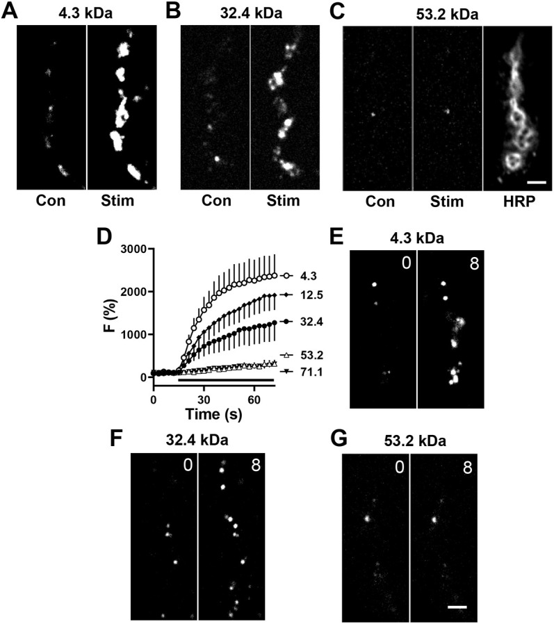
Permeability of PEG derivatives through DCV fusion pores. (A–C) Contrast-enhanced fluorescent images of type Ib boutons from Ok6-GAL4 UAS-Dilp2FAP larvae before (Con) and after 70 Hz stimulation (Stim) for 60 s in the presence of 4.3 kDa (A), 32.4 kDa (B) and 53.2 kDa (C) MG-PEG dyes, each at 1 µM. Anti-horseradish peroxidase immunofluorescence (HRP) is shown to indicate location of boutons. (D) Time-course of activity-evoked FAP responses in the presence of MG-PEG dyes normalized to initial labeling. Apparent protein sizes shown in symbol labels. Black bar, 70 Hz stimulation. 4.3 kDa: n=8 NMJs (one bouton each, four animals); 12.5 kDa n=7 NMJs (one bouton each, four animals); 32.4 kDa : n=5 NMJs (one bouton each, five animals); 53.2 kDa n=6 NMJs (one bouton each, five animals); 71.1 kDa n=6 NMJs (one bouton each, five animals). (E–G) Contrast-enhanced fluorescent images of Dilp2-FAP expressing boutons in the absence of Ca2+ after application of 4.3 kDa (E), 32.4 (F) and 53.2 kDa (G) MG-PEGs. Numbers on the images indicate time in minutes. Images in E–G representative of 10–12 experiments. Scale bars: 2 µm.
