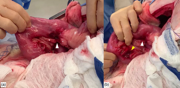
FIGURE 1.

Intraoperative images showing the two lesions. (a) The surgeon is retracting the proximal duodenum, showing the more orad perforation, indicated by a white arrowhead. The pylorus (P) and body of the pancreas (*) are indicated. (b) The surgeon is retracting the duodenum towards the duodenocolic ligament, the aborad perforation is indicated by a yellow arrowhead.
