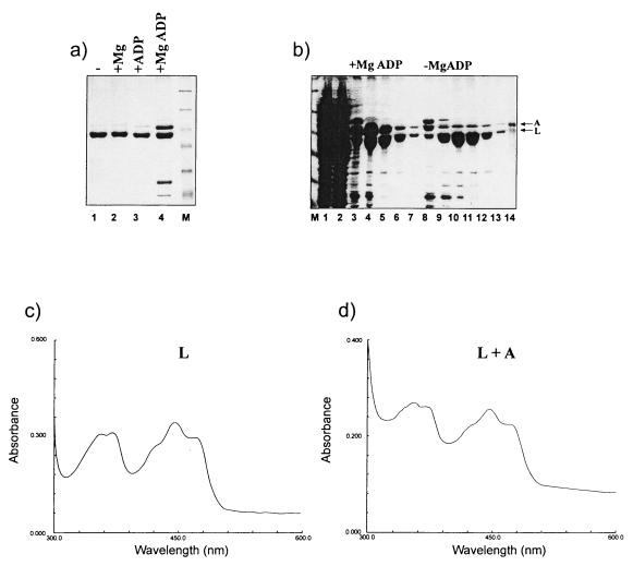
FIG. 5.
Isolation of the NIFL-NIFA complex directly from lysed cells. BL21(DE3)(pLysS) cells carrying plasmid pPR38 were grown as described in Materials and Methods. (a) The cell paste was resuspended in lysis buffer and divided into four aliquots. These were lysed and chromatographed as described in Materials and Methods with the following additions: lane 1, no nucleotide; lane 2, with 1 mM magnesium acetate; lane 3, with 1 mM ADP; lane 4, with 1 mM MgADP. Lanes 1 to 4, fractions containing protein which eluted with 0.5 M imidazole. Lane M contains molecular weight markers. (b) Cell paste was resuspended in lysis buffer containing 1 mM MgADP and was divided into two aliquots. These were applied to the nickel chelating column and washed with equilibration buffer containing 1 mM MgADP as described in Materials and Methods. Lane 1, cell supernatant; lane 2, nonbound protein from cell supernatant; lanes 3 to 7, NIFL-NIFA-containing fractions eluted with 0.5 M imidazole in the presence of 1 mM MgADP; lanes 8 to 13, fractions eluted with 0.5 M imidazole after washing the column in equilibration buffer without nucleotide to dissociate NIFA. (c and d) Absorbance spectra of oxidized NIFL and isolated NIFL-NIFA complex in the presence of 500 μM MgADP. The spectra were recorded with a Shimadzu MP2000 spectrophotometer with a 1-cm light path and 1-nm slit width.

