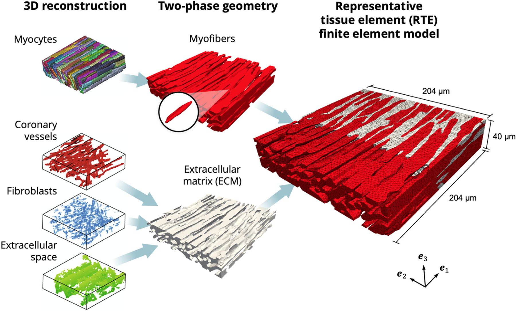
Fig. 3.

Finite element model developed from 3D reconstructions. Left: Geometry of myocytes, coronary vessels, fibroblasts, and extracellular space (Seidel et al., 2016). Center: Myocytes joined into the myofiber phase (red), with representative myocyte highlighted. Coronary vessels, fibroblasts, and extracellular space joined into the extracellular matrix (ECM) phase (gray). Right: Cross-section of RTE FE model showing myofiber elements embedded in ECM elements, with ECM elements removed to highlight myofiber interconnections. Coordinate axes {} indicate the image and laboratory axes.
