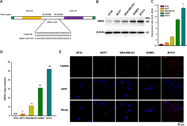
Fig. 1.
Uptake of TFO by different breast cancer cell lines. A. TFO is designed to bind to a polypurine sequence located in the exon of the HER2 gene. The HER2-11074 used in this article was designed to bind to a region between exon 23 and 24. B. Western blot analysis of HER2 protein levels in breast cancer cell lines with different gene copy numbers. C. Quantification indicated the level of HER2 in these cells. Data are presented as mean ± SEM and were analyzed by Tukey's post hoc test; Error bars represent SEM of the technical replicates (3 biological replicates with at least 2 technical replicates each). *, P = 0.03, BT474 compared with other cells; N = 3 independent experiments. D. Gene copy number characteristics of breast cancer cell lines. E. Confocal images show the nucleation of TFO in different breast cancer cells. The red fluorescence indicates the TAMRA-tagged TFO fragment (Scale bar = 50 μm)

