
Figure 3.
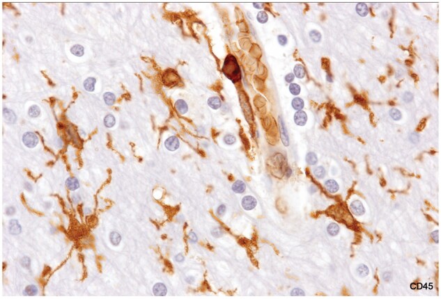
Reactive microglia, intact white matter. Cell bodies are enlarged, and processes are thickened. The myelinated tissue appears otherwise normal with normal-appearing oligodendrocytes. There is a monocyte in the wall of the blood vessel (Case 17, CD45, ×1020).