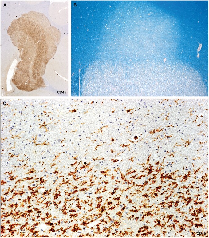
Figure 7.
A largely prephagocytic early plaque capped by an area of myelin pallor (A, B), largely preserved oligodendrocytes, and activated microglia (C). Immediately below this zone is a band of vacuolated myelin sheaths and oligodendrocyte loss. It was difficult to identify any LFB-positive phagocytes in the lesion and there were no MRP14-positive cells except in blood vessels (Case 2, A, CD45, ×4.5, B, Luxol fast blue, ×30, C, CD45, ×400).

