
Fig. 4.
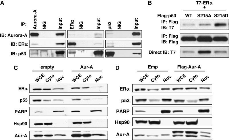
Aurora-A forms ternary complex with ERα and p53, and Aurora-A overexpression causes cytoplasmic sequesteration of them. a MCF7 cells were immunoprecipitated with indicated antibodies or normal IgG (NIG) followed by immunoblotting with indicated antibodies. Aliquots of the same whole cell lysates were directly immunoblotted with anti-Aurora-A antibody (bottom). b T7-tagged ERα was co-transfected with Flag-tagged p53 WT or SA or SD into MCF7 cells. Twenty-four hours after transfection, cells were subjected to immunoprecipitation with anti-Flag antibody followed by immunoblotting with indicated antibodies (top and middle). Whole cell lysates were directly immunoblotted with anti-T7 antibody (bottom). c Stable clone expressing empty vector and Aurora-A was fractionated into cytosolic (Cyto) and nuclear (Nuc) fractions and analyzed by immunoblotting with anti-ERα and p53 antibodies. The purity of cytosolic and nuclear fractions was confirmed by immunoblotting for anti-Hsp90 and anti-PARP antibodies, respectively. WCE whole cell lysates. d MCF7 cells transiently transfected with empty vector or flag-tagged Aurora-A for 24 h were fractionated as in c