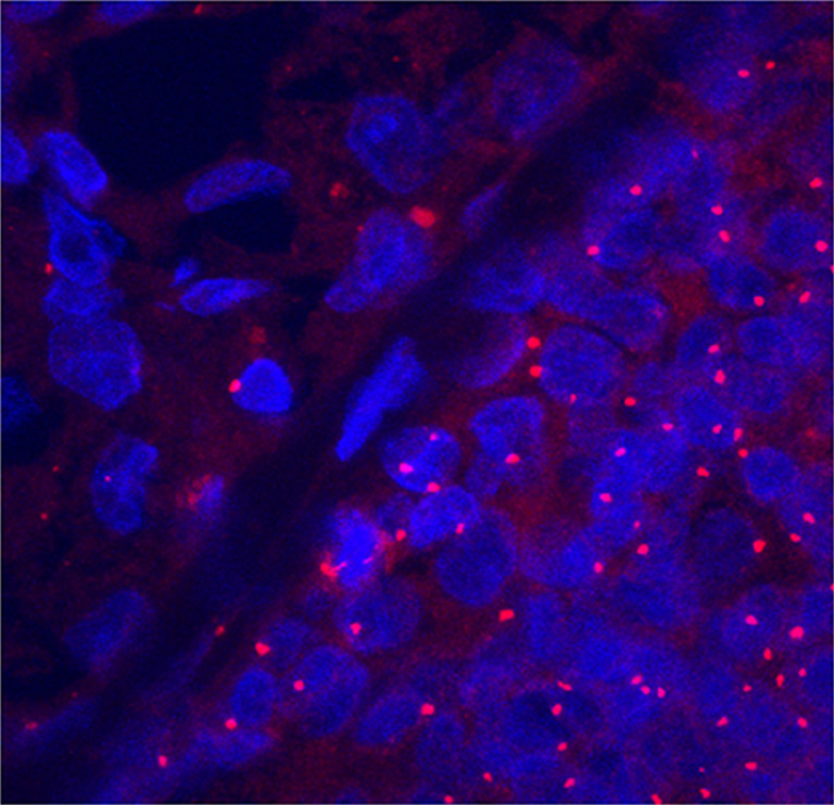
Fig. 1.

Centrosome amplification in E2-induced mammary tumors (MTs). Centrosomes and nuclei were observed by confocal microscopy of sections labeled with an antibody against γ-tubulin (red) and DNA dye Hoechst 33342 (blue) in areas corresponding to hematoxylin/eosin stained sections. In E2-induced MT cells (lower right), the centrosomes were consistently amplified in both size and number compared to adjacent normal hyperplasia (upper left).
