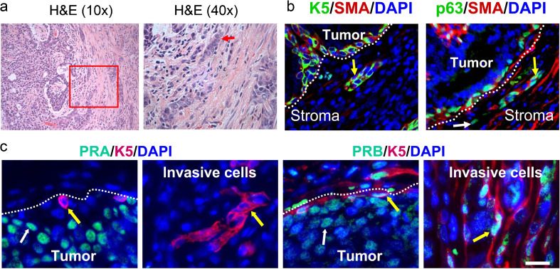
Fig. 3.
Invasive cells in the rat mammary tumors express basal/myoepithelial markers. Tumors were from carcinogen-treated ovary-intact (OI) or ovariectomized rats treated with estradiol alone (E) or estradiol plus progesterone (E + P). a H&E staining. Low-power image of an invasive tumor (×10, left panel). Inset indicates the area of the tumor at higher power (×40, right panel) with a cord of invading cells (red arrow). b Immunolabeling of invasive cells with anti-K5 (green) and anti-SMA (red) antibodies (left panel; yellow arrow indicates a basal/myoepithelial K5 + SMA + cell) or with anti-p63 (teal) and anti-SMA (red) antibodies (right panel; white arrow indicates a basal p63 + SMA cell, yellow arrow indicates a basal/myoepithelial p63 + SMA + cell). c Immunostaining of PR isoforms in luminal cells in the tumor body and in invasive basal/myoepithelial cells. Immunolabeling with anti-K5 (red) and anti-PRA (teal) antibodies (left two panels) or anti-PRB (teal) antibodies (right two panels). White arrows indicate PRA+ or PRB+ tumor luminal cells, yellow arrows indicate basal/myoepithelial cells. b, c White dotted lines indicate the margin of the tumor. Nuclei were counterstained with DAPI (blue). Scale bars, 50 μm

