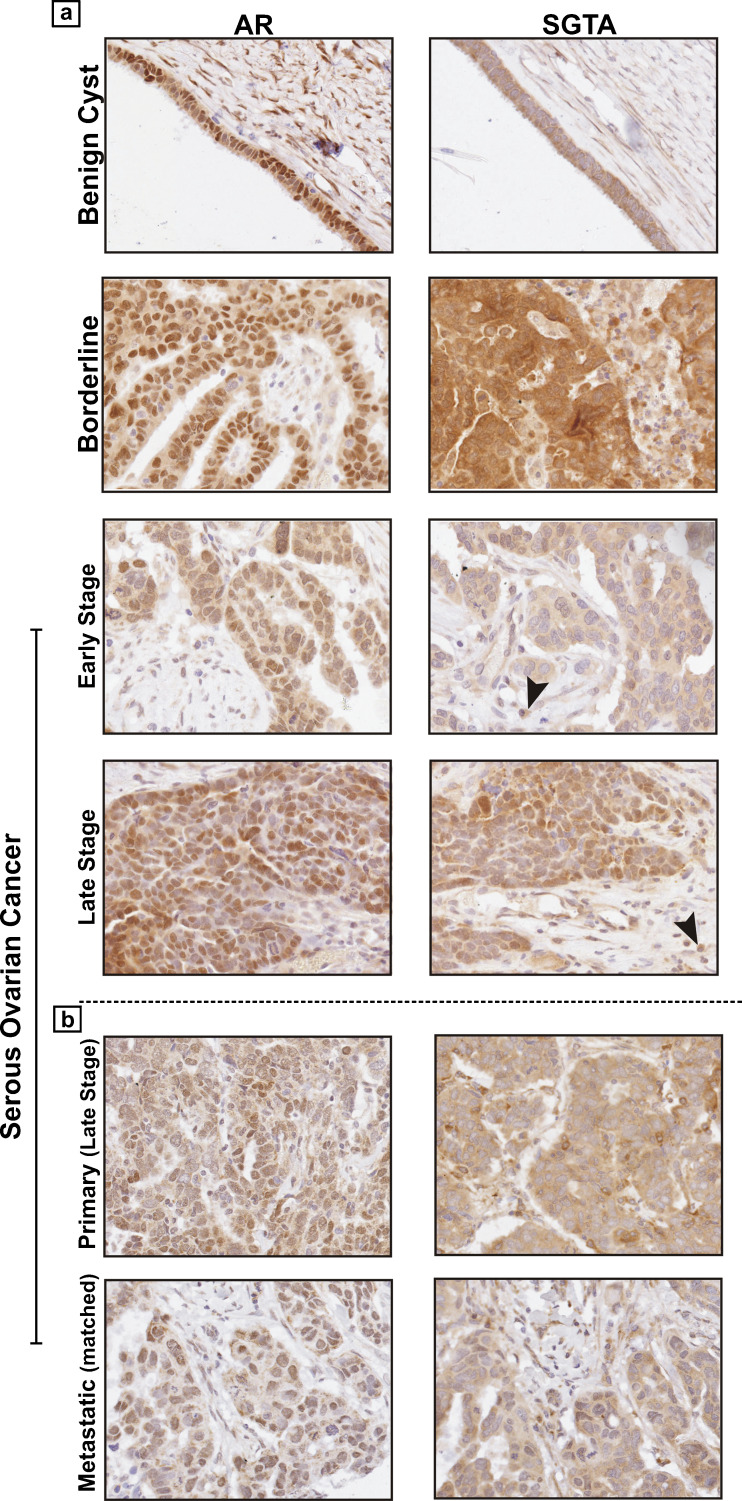
Fig. 2.
Immunohistochemical staining of AR and SGTA in serous epithelial benign and ovarian cancer samples. Images (×400 magnification) are representative examples from individual patient samples that correspond to AR (left) and SGTA (right) immunostaining in the epithelium of a malignant tumors (early and late stages), borderline tumors, and benign disease and b primary and matched metastatic tissues from an individual patient. Black arrowheads indicate epithelial cells that have invaded the stroma and are positive for SGTA in the cytoplasm

