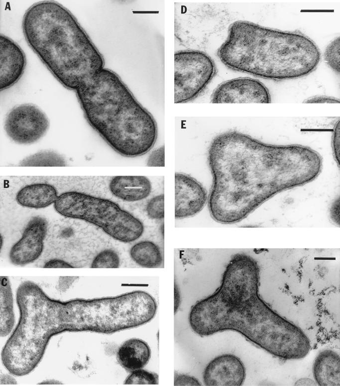
FIG. 2.
Different types of cell division revealed in electron micrographs of thin-sectioned cells. (A) Example of cell binary division with peptidoglycan layer and cytoplasmic membrane invagination. The nucleoid is seen as light zones. (B) Asymmetric constriction; (C) interesting type of cell division; (D through F) different stages of Y cell division. The nucleoids are distributed in three directions. Bars, 0.25 μm.

