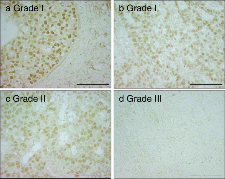
Fig. 3.
Immunohistochemistry of FOXA1 in breast cancer. The representative images of the immunohistochemical staining of breast cancer tissues with the anti-FOXA1 antibody are shown. Positive staining for FOXA1 was observed in the nuclei of the breast cancer cells. The FOXA1 immunoreactivities in grade III breast cancer were significantly lower than those in grade I breast cancer (5.83 vs. 2.38, P = 0.002). Bar, 100 μm

