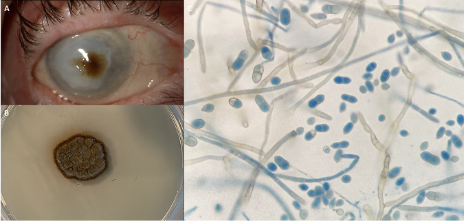
FIG 1.
(A) Right eye with therapeutic contact lens showing a brownish corneal infiltrate upon slit lamp examination. (B) Macroscopic view of the colony grown on SDA after 5 days of incubation at 30°C. (C) Microscopic view of lactophenol cotton blue mount, showing smooth, flexuous, pigmented, septate hyphae producing two-celled conidia with a small denticle.

