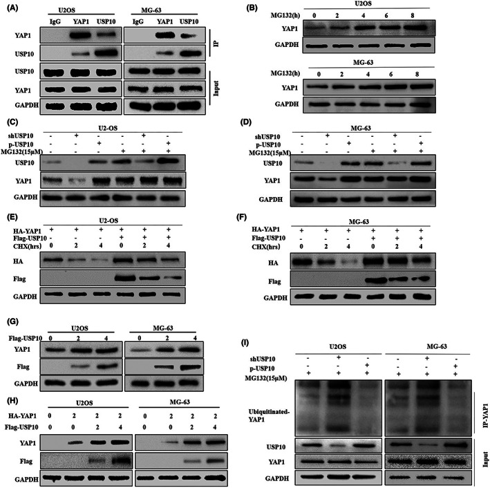
FIGURE 8.

USP10 stabilizes the expression of YAP1 by mediating its deubiquitination in OS cells. (A) The co‐IP results confirmed that USP10 and YAP1 interact directly in osteosarcoma cells. (B) The expression of YAP1 in U2OS and MG‐63 osteosarcoma cells treated with protease inhibitor MG132 (15 μM) was detected by western blotting. (C, D) Western blot results showed that the expression level of YAP1 did not change significantly after knockdown or upregulation of USP10 osteosarcoma cells treated with MG132. (E, F) Osteosarcoma cells U2‐OS and MG‐63 were transfected with the plasmid encoding HA‐YAP1, in the presence or absence of the Flag‐USP10 plasmid. It was then treated with CHX (20 μM), and finally, the interpretation of YAP1 was observed by western blotting. (G) Endogenous YAP1 expression levels were measured by transfecting OS cells with different doses of Flag‐USP10 plasmid, (H) U2‐OS and MG‐63 cells were transfected by plasmid encoding HA‐YAP1 or in combination with Flag‐USP10 plasmid. YAP1 expression was observed by an anti‐HA antibody. (I) Osteosarcoma cells were transfected with knockout/overexpressing USP10 plasmids and treated with MG132 (15 μM), and ubiquitinated YAP1 levels were assessed by western blotting with anti‐Ub antibody.
