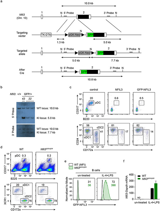
Extended Data Figure 1. Generation of Nfil3GFP fusion protein reporter mice.
a, Schematic diagrams of the mouse Nfil3 WT allele, the targeting vector and targeted allele. Filled and open boxes denote coding and noncoding exons of Nfil3, respectively. N indicate NdeI site. Triangles indicate loxP sequences. TK, thymidine kinase promoter; DTA, diphtheria toxin A; pGK-Neo, neomycin selection cassette. b, Southern blot analysis of Nfil3+/+ and Nfil3GFP/+. Genomic DNA was isolated from mouse liver, digested with NdeI, electrophoresed, and hybridized with Digoxigenin-labeled probes indicated in a. Southern blot with 5’ probe gave a 10.0 and a 5.0 kb band for WT and targeted allele. Southern blot with 3’ probe gave a 10.0 and a 7.7 kb band for WT and targeted allele respectively. Progeny from ES cell clone 22 were bred to CMV-Cre mice to remove the neomycin selection cassette, and used in the following study. c, Flow cytometric analysis showing pDCs and cDCs differentiated from WT CD117hi BM progenitors retrovirally expressing NFIL3 and GFP-NFIL3. The CD24+ CD172a− cDC1s and CD172a+ cDC2s are pre-gated as CD317− B220− MHCII+ CD11c+ cells. Data shown are one of two similar experiments. d, Representative flow plots showing pDCs and cDCs among live splenocytes from WT and Nfil3GFP/GFP mice, cDCs are pre-gated as CD317− B220− MHCII+ CD11c+ cells. Data shown are one of three similar experiments. e, Representative flow plots showing GFP-NFIL3 expression in B cells from Nfil3GFP/GFP mice. Whole splenocytes were cultured with medium or were stimulated with IL-4 (20 ng/mL) and LPS (5 μg/mL) for 24 h. B cells were pre-gated as CD19+ cells. Data shown are one of two similar experiments. f, Nfil3 transcripts, measured by RT-qPCR, in B cells sorted from e.

