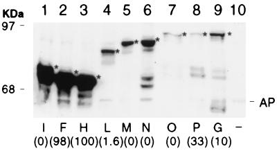
FIG. 5.
Detection of ComP-PhoA hybrid proteins in E. coli by Western blotting. Total cell extracts containing equal amounts of protein were separated by SDS-PAGE on a 10% gel and visualized by immunoblotting with monoclonal anti-alkaline phosphatase antibody. Fusion proteins are indicated by letters, and their positions are denoted by arrows. The numbers in parentheses represent alkaline phosphatase values normalized to that associated with fusion H (Table 2). A strain carrying the phoA vector with no comP insert was used as a negative control (−). The positions of molecular size standards (in kilodaltons) are shown, as is the position of mature alkaline phosphatase (AP).

