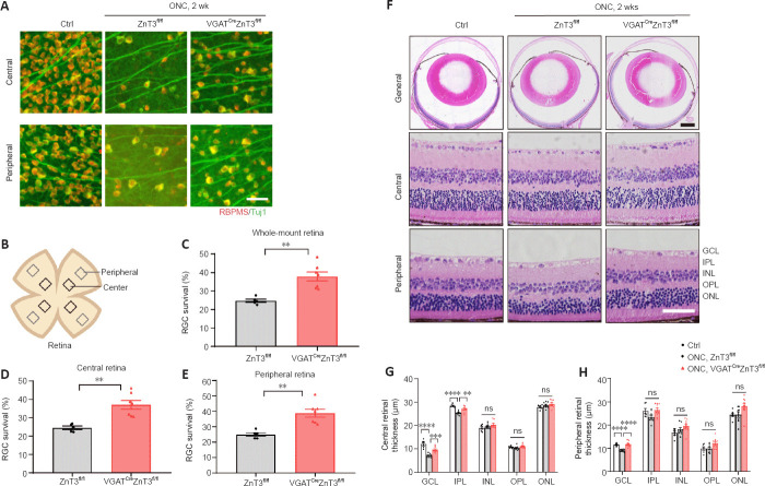
Figure 4.
Knockout of ZnT3 in ACs promotes RGC survival.
(A) Retinal whole-mounts immunostained for RNA binding protein with RBPMS (red) and Tuj1 (green) to visualize RGCs in the retinas of VGATCreZnT3fl/fl mice and the littermate control ZnT3fl/fl mice 2 weeks post-injury. RGC survival was increased in VGATCreZnT3fl/fl mice. Scale bar: 50 µm. (B) Schematic diagram of the whole-mount retina, showing the eight areas from the central and peripheral areas of the retina used for quantification. (C–E) Quantitative RGC counts in whole-mount, peripheral, and central retinal samples (n = 5, 7). Animals without ONC were used to be comparison group (n = 6). (F) Representative low- and high-power images of hematoxylin and eosin-stained cross sections. Compared with ZnT3fl/fl mice, VGATCreZnT3fl/fl mice showed attenuated retinal thinning. Scale bar: 50 µm for high-power images and 500 µm for low-power images. (G, H) Quantitative analyses of retinal thickness in the central and peripheral region of the retina (n = 6, 8, 10). Data are presented as mean ± SEM. **P < 0.01, ***P < 0.001, ****P < 0.0001 (unpaired two-tailed Student’s t-test (C–E) or one-way analysis of variance followed by Tukey’s multiple comparisons (G, H)). AC: Amacrine cell; Ctrl: control; GCL: ganglion cell layer; INL: inner nuclear layer; IPL: inner plexiform layer; ns: not significant; ONC: optic nerve crush; ONL: outer nuclear layer; OPL: outer plexiform layer; RBPMS: RNA binding protein, mRNA processing factor; RGC: retinal ganglion cell; VGAT: vesicular γ-aminobutyric acid transporter; ZnT3: zinc transporter 3.

