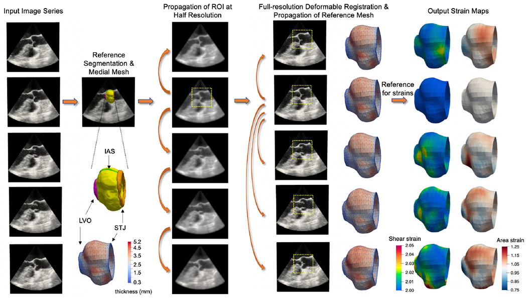
Figure 1:

The proposed semi-automated image segmentation, modeling, and strain analysis pipeline. A reference 3D volume is selected from the input image series and manually segmented. The aortic root is labeled in yellow, sinotubular junction (STJ) in orange, left ventricular outlet (LVO) in pink, and interatrial septum (IAS) in green. A medial mesh, with thickness shown in color, is generated from the reference segmentation. Consecutive pairs of images are then registered at half-resolution in order to propagate a masked region of the root to all frames in the series. Next, full-resolution deformable registration is performed between the reference frame and all other frames in order to propagate the reference medial model to all time points in the series. Maps of shear and area (dilatational) strain are computed from the series of medial models of the aortic root.
