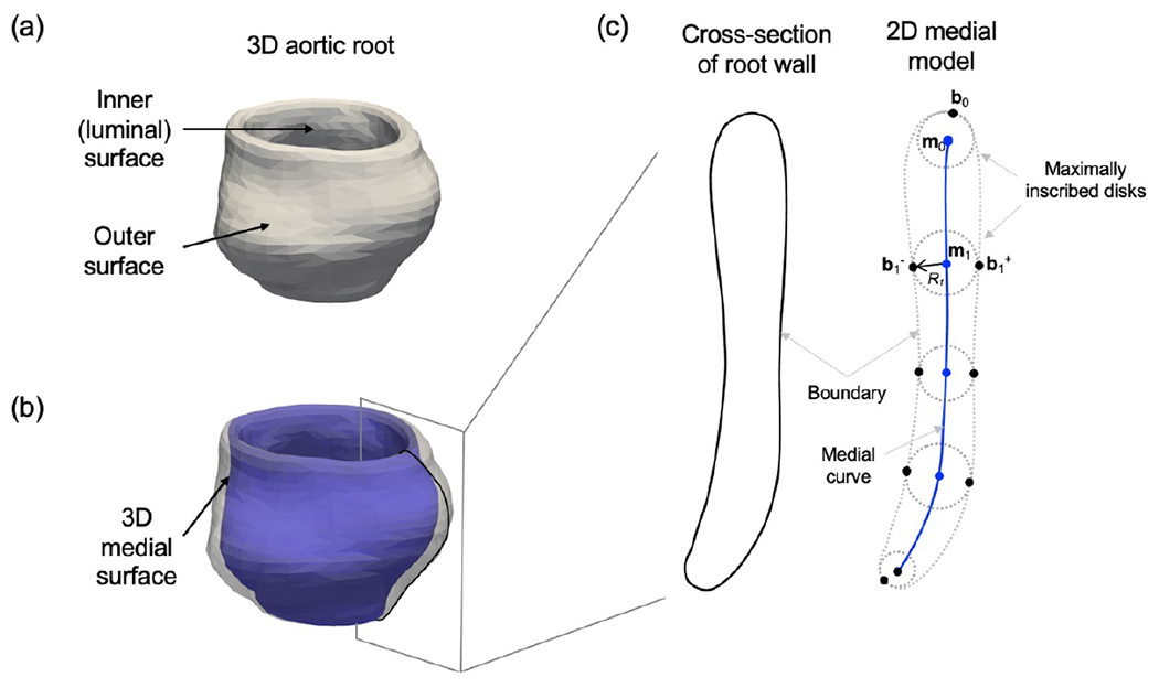
Figure 2:

Diagram of cross-sectional medial geometry. (a) Representative 3D aortic root wall with thin cylindrical geometry. (b) 3D aortic root wall shown with a translucent boundary. The medial surface (blue) passes midway between the luminal surface and outer surface of the root wall. (c) The 2D medial geometry of a cross-section of the root wall. The point m0 is the center of a maximally inscribed disk that is tangent to the root wall boundary at b0. The maximally inscribed disk centered at m1 has radius R1 and is tangent to the root wall surface at two medially linked boundary points: b1+ and b1−.
