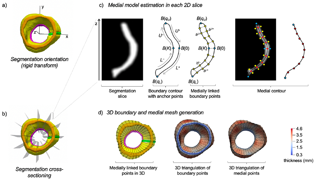
Figure 3:

Generation of a 3D medial mesh from an image segmentation of the aortic root. (A)The aortic root segmentation is rigidly transformed so that the outflow tract is vertically oriented along the z-axis and the interatrial septum (green) is aligned with the x-axis. (B) The oriented segmentation is rotationally cross-sectioned into long-axis slices of the root. (C) In each cross-section, a 2D non-branching medial axis is approximated using a surface resampling technique, wherein points on the medial axis are estimated as midpoints between medially linked boundary points on the root surface. (D) 3D triangulated surfaces of the root boundary and medial surface are generated by defining edges between nodes in neighboring cross-sections. Locally varying thickness of the aortic root is shown in color.
