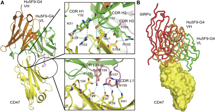
FIGURE 7.
Three-dimensional (3D) structure diagram of the complex of Hu5F9-G4 and CD47 showed that Hu5F9-G4 had competitive antagonism with SIRPα. The orange and green peptide chains are the heavy chain and light chain of Hu5F9-G4, respectively. The light chain variable region peptide is represented in pink. The yellow peptide chain represents CD47, while the red peptide chain represents SIRPα. (A) Three-dimensional structure of CD47-ECD and Hu5F9-G4 complex after crystallization (PDB 5IWL). All complementary determiners of Hu5F9-G4 heavy chain and light chain variable CDR1 and CDR2 rings help Hu5F9-G4 bind CD47-ECD epitopes. (B) Hu5F9-G4 and SIRPα share binding pockets, which can be observed by superposition of Hu5F9-G4/CD47-ECD and SIRPα/CD47-ECD structures (PDB ID 2JJS).

