Figure 2.
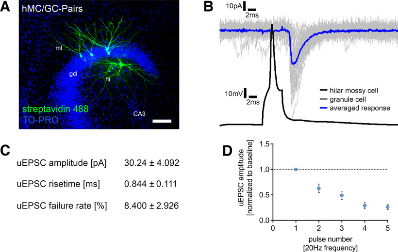
Single-cell characterization of the hilar mossy cell–dentate granule cell projection. A, Post hoc staining of paired recordings from hilar mossy cells (hMC) and dentate granule cells in the suprapyramidal blade of the dentate gyrus. TO-PRO nuclear stain was used to visualize cytoarchitecture. Scale bar, 100 µm. gcl, granule cell layer; ml, molecular layer; hi, hilar region. B, Action potentials were induced in hilar mossy cells (50 action potentials, black trace), and postsynaptic responses were recorded in dentate granule cells (gray traces, single sweeps; blue trace, averaged response). Connected pairs showed highly reliable inward currents on presynaptic stimulation. C, Summary table for single-cell characterization of excitatory inputs onto dentate granule cells, originating from hilar mossy cells (5 pairs in 21 tissue cultures). D, Short-term plasticity experiments revealed a depressive and depletive behavior of this connection following repetitive stimulation. Values represent mean ± SEM.

