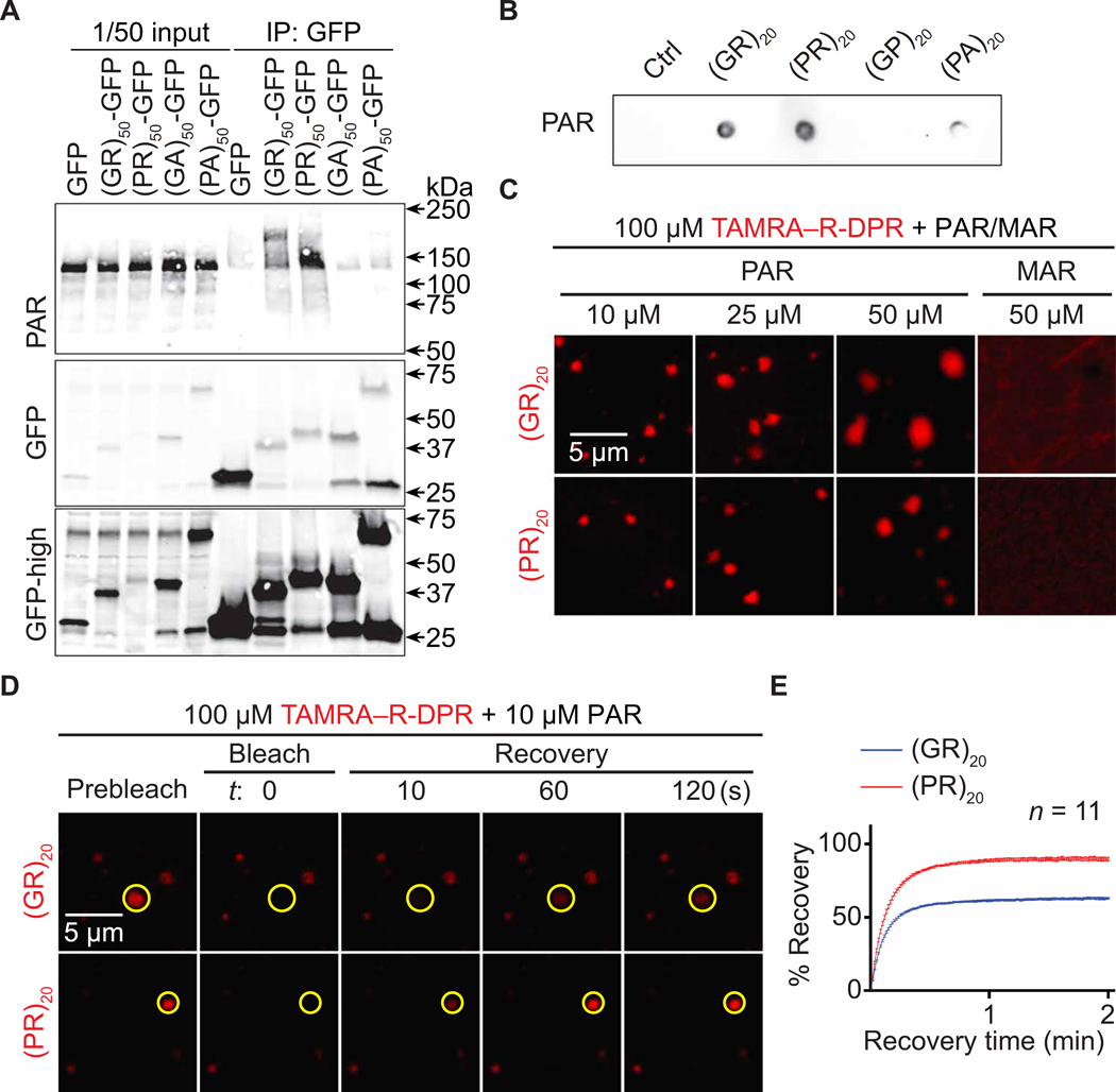
Fig. 3. PAR induces R-DPR condensation in vitro.
(A) Co-IP of PAR and DPRs in HEK293T cells. (B) Dot blot binding assays of PAR and DPRs. (C) TAMRA–R-DPR (red) mixed with PAR (10 to 50 μM MAR equivalent) or MAR. (D and E) FRAP assays on PAR and TAMRA–R-DPR (red) condensates. Yellow circles indicate bleached and analyzed areas. Total R-DPRs (1 μM) were labeled. Buffer for (C to E): 61.5 mM K2HPO4 and 38.5 mM KH2PO4

