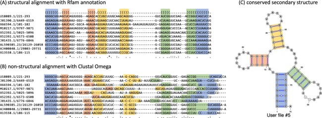
Figure 1.
Conserved secondary structures of transfer RNAs. (A) A multiple sequence alignment of 10 tRNAs extracted from the Rfam database is depicted, along with the annotated secondary structure. A string consisting of ‘.’, ‘(’ and ‘)’ at the top of the multiple alignment is the dot-bracket notation that represents the secondary structure. (B) A multiple sequence alignment based on sequence identity was calculated with Clustal Omega [8] for the 10 tRNAs. (C) The conserved secondary structure annotated by the Rfam database was visualized using VARNA [9]. Each colored region of base pairs corresponds to the acceptor stem (blue), D arm (red), anticodon arm (yellow) and T arm (green), respectively.

