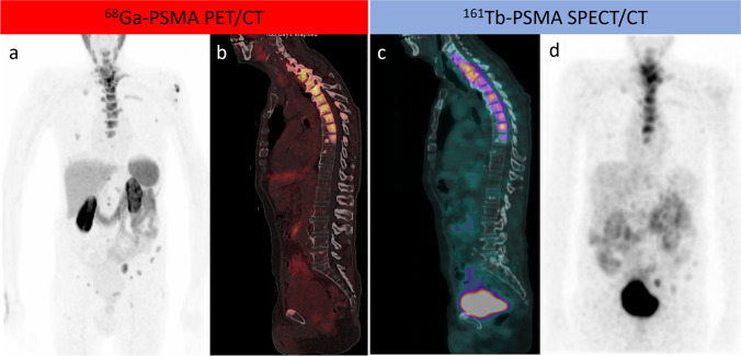
Fig. 1.
A patient with metastatic castration-resistant prostate cancer who progressed on hormonal therapy and was referred for PSMA-RLT. As shown in a maximum intensity projection (MIP) and b sagittal fused PET/CT, 68Ga-PSMA PET/CT revealed widespread PSMA-avid bone metastases that were extensive sclerotic in the thoracic spine. 161Tb-PSMA SPECT/CT 24 h after a 5.5 GBq theraputic dose of 161Tb-PSMA RLT demonstrated adequate localization of the 161Tb-PSMA in the metastatic bone deposits, which were extensively expressed in the thoracic spine as demonstrated by sagittal fused SPECT/CT and MIP (c, d)

