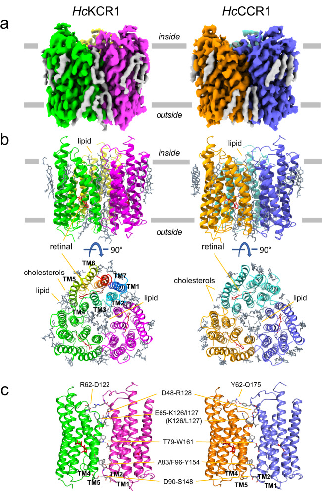
Fig. 2. Cryo-EM structures of HcKCR1 and HcCCR.
a Cryo-EM density maps of the HcKCR1 and HcCCR homotrimers viewed from the membrane plane. Protomers are colored in magenta, green, and yellow for HcKCR1, and in blue, orange, and turquoise for HcCCR, respectively. b Structure models viewed from the membrane plane (upper) and intracellular side (lower) with all-trans-retinal shown in red. In one protomer of HcKCR1 trimer helices are shown in different colors and labelled to show the order of transmembrane helices. c Interprotomer connecting residues observed in the trimeric configuration.

