Figure 2.
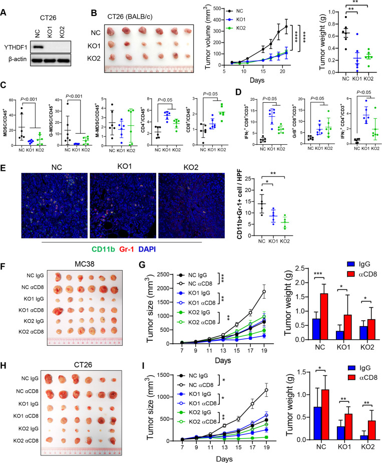
YTH N6-methyladenosine RNA binding protein 1 (Ythdf1) knockout induces antitumour immunity by reduction of myeloid-derived suppressor cell (MDSC) and increase of functional T cells in syngeneic tumours, an effect reversed by CD8+ T cell depletion. (A) Western blot validated Ythdf1 knockout in CT26 cells. (B) Representative image of CT26 syngeneic tumours with or without Ythdf1 Knockout (left). Knockout of Ythdf1 in CT26 cells inhibits tumour growth (middle) and tumour weight (right) in BALB/c mice. (C) MDSC, G-MDSC, M-MDSC, CD4+ T cells and CD8+ T cells from tumours in (B) were analysed by flow cytometry. (D) Flow cytometry analysis assessing the percentage of T cell functional markers interferon gamma (IFN-γ) and granzyme B (GrB) from tumours in (B). (E) Immunofluorescence identifying MDSCs in subcutaneous tumours from BALB/c injected with the indicated CT26 cells (n=5 each group). (F) MC38 NC or Ythdf1 knockout cells were implanted in C57BL/6 (n=6). Isotype control (IgG) or anti-mouse CD8 antibody (αCD8) were given at 200 µg/mouse on days 4, 7 and 9 post cell injection. Representative images of tumours from each group were shown. (G) Tumour volume (left) and weight (right) from tumours in (F). (H) CT26 NC or Ythdf1 knockout cells were implanted in BALB/c (n=6). Isotype control (IgG) or αCD8 were given at 200 µg/mouse on days 4, 7 and 9 post cell injection. Representative images of tumours from each group were shown. (I) Tumour volume (left) and weight (right) from tumours in (H) (*p<0.05; **p<0.01; ***p<0.001; ****p<0.0001) (two tailed t-test (B, C, D, E, G, H), two-way analysis of variance test (D, G, I)). NC: cells with control sgRNA. KO1: cells with YTHDF1 sgRNA #1. KO2: cells with YTHDF1 sgRNA #2.

