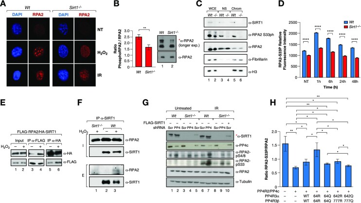
Figure 5.
SIRT1 regulates RPA2 phosphorylation through PP4. (A) IF analysis of γH2Ax and RPA2 foci formation in Wt or Sirt1−/− MEFs under or not oxidative stress (2 mM H2O2 for 1 h), or ionizing irradiation (IR 30 min 7.5 Gy). (B) Western-blot analysis of the levels of the total pRPA2 in Wt and Sirt1−/− MEFs previously treated with 7.5 Gy IR. Quantification of n = 3 experiments and representative experiment are shown. The shifted phosphorylated form of RPA2 is detected in the longer exposure blots and is marked with *. Two-tail analysis (**P< 0.01). (C) Western blot of SIRT1, RPA2 (total and S33ph) in whole cell extracts (WCE), nuclear soluble fraction (NS) and chromatin insoluble fraction (Chrom) of Wt or Sirt1−/− U2OS cells previously treated with 2 mM H2O2 for 1 h. The levels of fibrillarin and histone H3 were also analyzed as controls of NS and Chrom fractions, respectively. (D) IF Time-course experiment of RPA2-S33P performed as in 3A. A similar experiment is shown by western blot in Figure S4B. (E) Co-IP between FLAG-RPA2 and HA-SIRT1 using FLAG and HA resin under untreated condition or oxidative stress (2 mM H2O2 for 1 h) in HeLa cells. (F) Endogenous IP with α-SIRT1 antibodies of RPA2 and SIRT1 from whole cell extracts of U2OS cells previously treated with 2 mM H2O2 for 1 h. Sirt1−/− extracts were used as a negative control. (G) Levels of the indicated proteins and RPA2 marks upon shRNA-driven downregulation or not of PP4c in Wt and Sirt1−/− MEFs untreated or irradiated with IR (7.5 Gy). Lanes 5 and 10, SIRT1 expression was re-introduced in Sirt1−/− (indicated with *). (H) Similar experiment as in 3D with RPA2-S33P levels.

