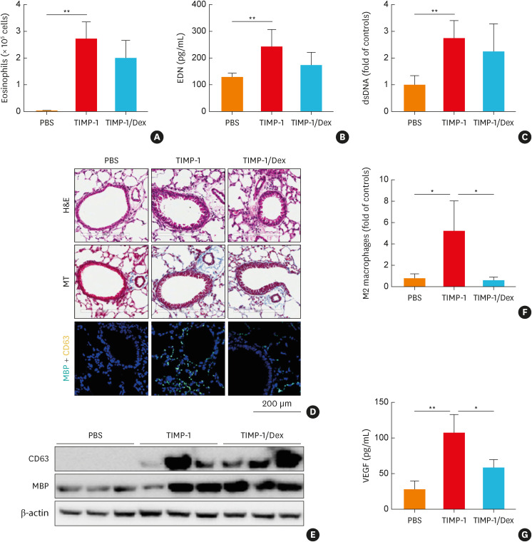
Fig. 5. The effects of TIMP-1 on airway inflammation in vivo. (A) Eosinophils in the BALF. The levels of (B) EDN and (C) dsDNA in the BALF. (D) Lung histology stained with H&E and MT staining. Immunofluorescence staining for DAPI (nuclear, blue), MBP (turquoise), and CD63 (yellow). Scale bar, 200 µm. (E) The expressions of CD63 and MBP in the lung tissues. (F) M2 macrophage counts in the lung tissues were obtained by flow cytometry. (G) The levels of VEGF in the BALF (n = 5 for each group). Data are presented as mean ± standard deviation.
BALF, bronchoalveolar lavage fluid; DAPI, 4’,6-diamidino-2-phenylindole; Dex, dexamethasone; EDN, eosinophil-derived neurotoxin; H&E, hematoxylin and eosin; MT, Masson’s trichrome; MBP, major basic protein; TIMP-1, tissue inhibitor of metalloproteinase-1; VEGF, vascular endothelial growth factor; PBS, phosphate buffered saline.
*P < 0.05 and **P < 0.01 by one-way analysis of variance with the Bonferroni post hoc test.

